Vestibular Rehab: BPPV Diagnosis and the Epley Maneuver
Learn how to diagnose the common posterior-canal variant of Benign paroxysmal positional vertigo (BPPV) by means of the Dix-Hallpike test and its treatment with the Epley maneuver, set out sequentially, together with the supporting evidence and the relevant safety considerations.

Benign paroxysmal positional vertigo (BPPV) is the most common cause of dizziness. An accurate diagnosis and a competently executed repositioning maneuver can resolve symptoms within a single consultation. This article describes the diagnosis of the common posterior-canal variant by means of the Dix-Hallpike test and its treatment with the Epley maneuver, set out sequentially, together with the supporting evidence and the relevant safety considerations.
What Is BPPV (Benign Paroxysmal Positional Vertigo)?
BPPV is a mechanical disorder of the inner ear. Otoconia (minute calcium carbonate crystals that ordinarily reside in the utricle) become dislodged and migrate into one of the semicircular canals. When the head moves, these displaced crystals induce abnormal fluid movement within the canal, producing a brief but intense illusion of rotation. Its principal features are as follows:
- Vertigo is provoked by changes in position, including turning over in bed, lying down, looking upward, or bending forward.
- Episodes are brief, typically lasting less than a minute, and subside once the head is still.
- The posterior semicircular canal is implicated in the substantial majority of cases, which is why the Dix-Hallpike test and the Epley maneuver constitute the core competencies.
- The condition is common and recurrent, and its prevalence rises with age. It is also associated with an increased risk of falls in older adults, and is therefore far from inconsequential.
The clinical interest of BPPV lies in the disparity between how alarming it feels and how amenable to treatment it is. Patients frequently present in considerable distress, convinced that something grave is occurring, and have sometimes been commenced on vestibular sedatives that fail to address the underlying cause. For many, a confident diagnosis followed by a maneuver that resolves the problem within minutes is transformative. This places the two core competencies described below, including accurate positional testing and correct repositioning, among the highest-value techniques a clinician can possess.
How Is BPPV Diagnosed? The Dix-Hallpike Test
The Dix-Hallpike test is the reference standard for diagnosing posterior-canal BPPV. It deliberately moves the suspected canal into a position that provokes the characteristic nystagmus and vertigo.
- Seat the patient on the couch, positioned such that, on lying back, the head will extend slightly beyond the edge. Explain that the test may briefly provoke their dizziness.
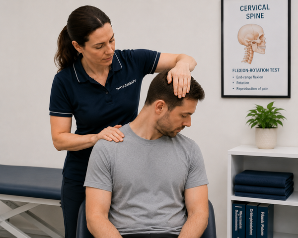
- Turn the patient’s head 45 degrees toward the side being tested.
- Maintaining the head in rotation, swiftly assist the patient to lie back so that the head is extended approximately 20 degrees below the horizontal. Support the head throughout.
- Observe the eyes for 30 to 60 seconds. A positive test produces a short latency followed by an upbeat and torsional nystagmus, accompanied by the patient’s familiar vertigo.
- Return the patient slowly to sitting, observe for reversal nystagmus, and then repeat on the opposite side.
- The side that provokes the nystagmus and vertigo is the affected side. The upbeating, torsional pattern confirms posterior-canal involvement. A purely horizontal nystagmus suggests lateral-canal BPPV, which requires a different test and a different maneuver.
How to Distinguish BPPV From Other Causes of Dizziness?
Dizziness is a broad presenting complaint, and BPPV is only one cause among many. The history accomplishes most of the differentiation before the patient is ever positioned. Three questions are particularly discriminating:
- Is it genuinely positional? BPPV is provoked by changes in head position relative to gravity, not simply by standing. Dizziness occurring only on standing points more toward orthostatic causes.
- How long does each episode last? BPPV episodes are brief, subsiding within a minute once the head is still. Vertigo persisting for hours suggests vestibular neuritis or Ménière’s disease, while constant unsteadiness suggests a different cause again.
- Are there auditory or neurological symptoms? Hearing loss or tinnitus directs attention away from BPPV and toward other inner-ear pathology. Neurological symptoms indicate a central cause.
- A characteristic history of brief, position-triggered rotation, with no auditory or neurological features, is highly suggestive of BPPV and establishes the Dix-Hallpike test as the confirmatory step.
When to Pause and Reconsider: Red Flags in Positional Vertigo
Most positional vertigo is benign; however, a small number of features should arrest treatment and prompt further assessment:
- Nystagmus that is purely vertical, purely torsional, without the typical pattern, direction-changing, or non-fatiguing, each of which raises the possibility of a central cause.
- Associated neurological symptoms, severe headache, or other red flags indicate a central rather than a peripheral problem.
- Caution with the positioning itself in patients with significant cervical disease, vascular concerns, or restricted neck range of movement; the technique or position should be modified as required.
How to Perform the Epley Maneuver: Step by Step
The Epley canalith repositioning maneuver guides the displaced crystals out of the posterior canal and back into the utricle, where they no longer provoke symptoms. The sequence below treats a right-sided BPPV; it should be mirrored for the left.
- Begin in the Dix-Hallpike position that provoked the symptoms: head turned 45 degrees toward the affected (right) side, lying back with the head extended. Hold until the nystagmus subsides, approximately 30 seconds or until symptoms abate.
- Without raising the head, rotate it 90 degrees toward the opposite (left) side, so that it is now turned 45 degrees to the left. Hold for approximately 30 seconds.
- Roll the patient onto the left shoulder while turning the head a further 90 degrees, so that they are now facing the floor at roughly 45 degrees. Hold for approximately 30 seconds.
- Assist the patient to sit up sideways, keeping the head turned, then return the head to neutral and tuck the chin slightly. The maneuver is complete.
- Each position is held until the provoked nystagmus and vertigo subside, typically about 30 seconds. The entire sequence requires only a few minutes. It is common practice to repeat the Dix-Hallpike test afterward to confirm resolution.
Setting Patient Expectations and Alternatives to the Epley Maneuver
Before commencing, advise the patient that the maneuver may briefly reproduce their vertigo and, occasionally, nausea, as the crystals move. This is expected and short-lived, and forewarning the patient substantially improves tolerance and cooperation. Several further practical points apply:
- Where the patient cannot tolerate the positions or has neck or back limitations, the Semont liberatory maneuver is a recognised alternative for posterior-canal BPPV.
- For patients who experience frequent recurrence, a home exercise such as the Brandt-Daroff exercise may be taught as a self-management option, although it is generally less effective than a clinician-performed repositioning maneuver.
- A single maneuver resolves many cases, but a proportion require a second or third attempt within the same consultation or at follow-up. Repetition is normal and does not indicate failure.
Does the Epley Maneuver Work? What the Evidence Shows
The Epley maneuver is strongly supported by the evidence. A review of the Epley maneuver for posterior-canal BPPV found it to be a safe and effective treatment, yielding significantly higher rates of both symptom resolution and conversion of a positive Dix-Hallpike test to negative when compared with sham or control. The 2017 guideline likewise recommends canalith repositioning as the first-line treatment.
Two practical implications follow:
- Vestibular suppressant medications are not recommended for routine BPPV; they do not address the mechanical cause and may prolong recovery. The guideline specifically discourages their routine use.
- Routine imaging is not indicated for a typical BPPV presentation and incurs cost without benefit.
BPPV Aftercare, Recurrence, and Follow-Up
A number of points complete the management picture:
- Post-maneuver positional restrictions were formerly routine, but the evidence does not support imposing strict ones; they are not necessary for a successful outcome.
- Reassess within a few weeks to confirm resolution. Should symptoms persist, repeat the maneuver, which frequently requires more than one attempt.
- Recurrence is common over the years, so the patient should be taught to recognise it and informed that re-treatment is straightforward.
- Should a patient remain symptomatic despite correctly performed repositioning, or should the presentation prove atypical, refer for specialist vestibular assessment.
KineticFlow For BPPV Assessment
KineticFlow helps you:
- Record the diagnostic findings: The affected side, nystagmus pattern and Dix-Hallpike result are documented, so the diagnosis is clear, and the right maneuver is chosen.
- Track resolution across visits: Pre- and post-treatment status and retest results are stored to show whether the maneuver worked.
- Flag atypical or persistent cases: A documented lack of response or atypical features prompts timely onward referral.
- Manage the recurrence pattern: Because BPPV recurs, the record lets each episode build on the last rather than starting fresh.
Try KineticFlow for your next patient assessment!
References
https://journals.sagepub.com/doi/full/10.1177/0194599816689667
https://journals.sagepub.com/doi/full/10.1177/0194599816689660
https://pubmed.ncbi.nlm.nih.gov/28248609/
https://www.cochranelibrary.com/cdsr/doi/10.1002/14651858.CD003162.pub3/full